23 Feb Doctors use 3D printing to correct Tessier Facial Cleft
Almost two-year Violet Pietrok was born with a rare condition called a Tessier Cleft, which meant that her bones didn’t fuse together prenatally. This left her with eyes so far apart that she couldn’t see properly, and her nose had no cartileage.
While Dr. John Meara of the Boston Children’s Hospital wanted to help Violet, the uniqueness of her condition meant that surgery could be extremely difficult – after all, no two skulls are quite the same, and reconstruction posed a challenge.
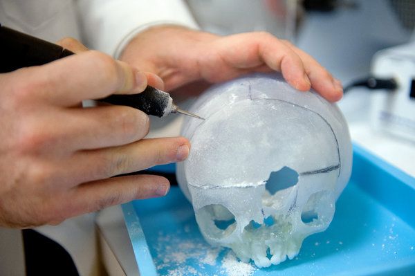
With the use of a 3D printer, Dr. Meara and his colleague Dr. Peter Weinstock were able to produce 3D models of Violet’s skull, using data from magnetic resonance imaging (MRI)’s. The printed skulls gave Dr. Meara the ability to practice on four different skull models, which allowed him to develop the best possible plan for Violet’s surgery ahead of time.
Dr. Weinstock, the director of the Pediatric Simulator Program at Boston Children’s, sees 3D models as part of a larger program to improve surgical craft. The ability to practice with models, he said, develops team communication, trust, and confidence before attempting extremely complex operations, and can shorten patients’ time under anesthesia.
Dr. Meara was able to move Violet’s eyes closer together and eliminate a large hole in her forehead. Due to the nature of her condition she will have to have more surgery as she grows, but 3D models of her skull have made the first steps that much easier.
No Comments